 From DNA to messenger RNA: transcription
 Transcription is an RNA biosynthesis that, like that of DNA, relies on base complementarity. A strand of DNA serves as a template. Transcription takes place in the nucleus. It consists of copying coded information contained in the DNA molecule into coded information contained in a messenger RNA molecule.
Unlike replication, which involves the entire genome at each cycle, the transcription program is not fixed: only small portions of the genome are transcribed at a given time in the life of the cell and these portions vary according to the development, environment etc …
Transcription, therefore, begins at a precise point of the DNA to end at an equally precise point, the space between the two constitutes a unit of transcription, a concept close to the cistron but not all exactly the same.
a single strand of DNA is transcribed, ie serves as a model for the polymerization of ribonucleotides. Indeed, a single strand of DNA in a given location has a meaning in terms of protein, which is why we generally write a DNA sequence in the form of a succession of bases from 5 ‘to 3’. In this convention, we actually represent the strand that has the code (coding strand) but it is the other strand (3 ‘- 5’ in our convention) that is transcribed. The result is an RNA molecule whose 5 ‘3’ orientation matches the NH2 - COOH orientation of the protein. The reading of the code (the translation) is done in the same direction as the transcription.
Note: All the transcription product does not correspond to the code of the protein, on the 5 '(upstream) side a guide or “leader” sequence allows the attachment of the messenger RNA to the ribosome.
Transcription is provided by an RNA polymerase that uses single-stranded DNA (as for replication, local denaturation of the DNA molecule is necessary) but it polymerizes ribonucleotides opposite deoxyribonucleotides.
Many protein factors are also involved in ensuring initiation, elongation and termination, but they are not the same as those involved in the replication.
Work of RNA Polymerase
 Transcription begins with the opening and unwinding of a portion of the DNA double helix molecule. As it progresses along with the DNA, the enzymatic complex that constitutes the RNA polymerase incorporates nucleotides present in the cell medium. This incorporation takes place by the complementarity of the nitrogenous bases with one of the strands of the DNA molecule: adenine (A) is placed opposite thymine (T) and uracil (U) is placed opposite adenine (A), cytosine (C) is placed opposite guanine (G) and vice versa.
The strand of messenger RNA thus synthesized is complementary to the strand of transcribed DNA. The information contained in the messenger RNA is identical to that of the non-transcribed DNA strand. The uracil nucleotide (U) occupies the place of the thymine nucleotide (T) in DNA in RNA.
RNA polymerase always moves in the same direction on one strand and in the opposite direction on the other strand. It is detached at a site with particular characteristics signalling the end of the gene. Then, when the RNA polymerase is detached, the two DNA strands join together again as the enzyme progresses, and find themselves as they were before RNA synthesis.
An amplification process
RNA is produced very quickly and in large quantities: several RNA polymerase molecules simultaneously transcribe the same gene.
Several genes can be transcribed simultaneously in the nucleus of the same cell.
Steps of Transcription
 Pre-initiation
 The promoter is a sequence of around one hundred nucleotides located in the regulatory region and designating the start of transcription. It is located upstream from the initiation site and carries sequence elements recognized by RNA polymerase and determining the direction of transcription.
The promoter is made up of short sequences conserved from one transcription unit to another and called consensus sequences
The intrinsic strength (or efficiency) of a promoter is defined by the relative number of initiation per unit time (rate of transcription). It depends on the proportion of the A-T base pairs relative to the CG base pairs, on the position of the sequences at -35 and at -10, in fact, the closer the consensus sequences are to the initiation site, the more the promoter will be. strong and ultimately strong RNA polymerase interaction with the promoter.
The promoter acts on the transcription of the DNA segment which is adjacent to it on the same chromosome, the promoter is said to be “cis” active. The promoter is not active on the coding sequences located elsewhere on the chromosome, in this case, we would then speak of the qualifier “ trans ”. The cis is opposed to the trans.
The affinity of RNA polymerase for DNA depends on the form of the enzyme: the core enzyme has a weak and non-specific affinity, the holoenzyme has a very strong and specific affinity for the promoter. We can note that the sigma σ subunit in the free state does not bind to DNA. The β 'subunit being basic and the DNA being acidic, it will be this which will facilitate the interaction of the complex with the promoter.
The sigma σ subunit, therefore, allows specific recognition of the promoter by RNA polymerase and decreases the affinity of the enzyme for non-promoter regions. It acts cyclically, indeed after the initiation made, the sigma factor is detached to be recycled and reused for other initiations of genes.
The RNA polymerase causes the denaturation of the two strands of DNA on 14 pairs of nucleotides, we speak of an open complex that further increases the affinity of the enzyme for the double helix.
 Initiation
 corresponds to the synthesis of the first phosphodiester bond produced by the β subunit which corresponds to the catalytic subunit of RNA polymerase.
The interaction of this subunit is inhibited by rifampicin which thus irreversibly inhibits DNA transcription, as is the case with tuberculosis. A mutation in SU β induces the appearance of bacterial strains resistant to rifampicin.
The flow of the first stages of transcription is, therefore:
- nonspecific binding of the holoenzyme.
- formation of a closed complex at the promoter level
- formation of the open complex (unwinding over 14 nucleotides)
- Placement of the first nucleotide (very often A or G)
- Elongation of 4 to 5 nucleotides
- Detachment of the sigma factor, after the transcription of the first 4-5 nucleotides.
 Elongation
 corresponds to the displacement of the transcription bubble along the DNA molecule. The mismatched region is then 70 base pairs. During transcription, the RNA forms a short pairing with the matrix strand of DNA forming a hybrid DNA-RNA helix on about ten base pairs.
Elongation is inhibited by aminoglycosides or amino glucosides.
 Termination
 occurs when the enzyme arrives at a specific sequence called a terminator.
The terminator is presented in the form of a palindrome (cf. lexicon) which can be perfect or imperfect. This palindrome results in sequence complementarity at the mRNA level which allows the establishment of a hairpin (or stem-loop) structure which is an intra-chain pairing that destabilizes the RNA polymerase up to dissociation.
It can be facilitated by a factor rho ρ depending on the sequence of the terminator, we thus highlight independent rho terminators (about 2/3) and rho dependent terminators (about 1/3):
For independent rho terminators, we find a hairpin structure rich in GC base pairs, followed by a poly-U sequence of about 6 nucleotides allowing easier dissociation of the DNA-RNA hybrid.
For the rho dependent terminators, we find a shorter hairpin structure that is not rich in GC base pairs and which is not followed by a poly-U sequence. There is therefore a need for the rho factor which has an affinity for RNA short of synthesis, traversing it from 5 ‘to 3’ until the RNA polymerase is found. The rho factor is ATP dependent, the hydrolysis of which will allow the dissociation of the complex.
 Maturation of primary transcripts
The primary transcript corresponds to immature RNA which requires processing in the form of cleavages or base modifications. This maturation is not obligatory. After maturation, mature RNA is obtained. We can take a few examples such as the maturation of the primary transcript giving the rRNAs (45S primary transcript) or the tRNAs by ribonucleases, or the maturation of the primary transcript encoding the mRNAs. Generally, there are few modifications for mRNAs; many mRNAs are translated into proteins while they are still being transcribed (Warning: there is no nucleus in prokaryotes).
The primary transcript codes either for a product, we speak of monocistronic RNA or several products, we will then speak of polycistronic RNA ( cf. beginning of the course, generalities ).
From RNA to protein: translation
 In the cell, translation takes place at the level of ribosomes: organelles made up of ribosomal RNA (rRNA) and proteins. Ribosomes are made up of two distinct subunits see figures 15 a and b, a large subunit and a small subunit, they are visible under an electron microscope.
The other actors in the translation are the transfer RNAs (tRNAs see FIG. 7) which carry the amino acids and the messenger RNA (mRNA) itself the sequence of which can be cut into codon from the AUG translation initiation codon.
In eukaryotes, ribosomes are either free in the cytoplasm or linked to the endoplasmic reticulum (this is called agranular endoplasmic reticulum) or to the nuclear membrane and translation takes place in the cytoplasm.
In prokaryotes, transcription and translation are simultaneous whereas in eukaryotes translation takes place in the cytoplasm from mature mRNA.
The messenger RNA produced in the nucleus passes into the cytoplasm to be translated there: this is the synthesis of proteins.
Stages of Translation
1. Initiation : The ribosome , a small cytoplasmic organelle (“protein assembly workshop”) begins reading the gene whose sequence is copied onto messenger RNA. It recognizes a place on the molecule called an initiator codon . Each ribosome is made up of two subunits: the small subunit carries a messenger RNA reading site and the large subunit has a catalytic site. A ribosome therefore behaves like an enzyme characterized by an active site. It catalyzes the amino acid polymerization of the protein that is being formed.
2. Elongation: The relative displacement of the ribosome and of the messenger RNA is accompanied by the progressive lengthening of the polypeptide chain; to each triplet of nucleotides of the messenger RNA corresponds a precise amino acid which is incorporated into the polypeptide chain being formed. The correspondence between triplets of messenger RNA nucleotides and amino acids is carried out according to the principles of the genetic code. The support for each of 20 amino acids is performed through RNA t (transfer RNA) specific part of which is fixed on each codon. peptide bond allows the attachment of each new amino acid (provided by an RNA transfer) to the last amino acid of the chain being extended. The new amino acid arriving in turn temporarily becomes the last.
3. Termination: The ribosome arrives on one of the three “stop” or “nonsense” codons, codon to which no amino acid corresponds. The dissociation between the messenger RNA and the terminated polypeptide chain then takes place.
The actors of translation
The actors of translation are messenger RNA (mRNA), transfer RNA (tRNA), ribosomes, amino acids, amino-acyl tRNA synthetases, Mg 2+, GTP and ATP.
1) Ribosomes
Ribosomes are made up of ribosomal RNA (rRNA) and proteins and are structured in the form of two subunits in either prokaryotes or eukaryotes ( see also general chapter ). Their size is defined in Svedberg units.
- Prokaryotic ribosomes (70S) consist of a small 30S subunit and a large 50S subunit.
- The 30S subunit consists of a 16S rRNA (1541 nucleotides) and 21 proteins.
- The 50S subunit consists of 23S (2904 nucleotides) and 5S (120 nucleotides) rRNAs as well as 32 proteins.
- Eukaryotic ribosomes (the 80S) consist of a small 40S subunit and a large 60S subunit.
- The 40S subunit is made up of an 18S rRNA and 33 proteins.
- The 60S subunit consists of 28S, 5.8S and 5S rRNAs as well as 49 proteins.
Schematic topography of the bacterial ribosome:
The bacterial ribosome has specific sites:
- Site A: (= Amino-Acid or Acceptor site) binding of amino acids.
- Site P: (= Peptide or Donor site) binding off-Met.
- Site E: (= Exit site) exit of the transfer RNA.
- EF-G site: present at the level of the large sub-unit.
- EF-Tu site: present at the level of the small sub-unit.
Attention in eukaryotes the first amino acid is methionine and not the f-Met present in prokaryotes.
The chaining of ribosomes on mRNA forms the polysome, it makes it possible to increase the efficiency of translation. The minimum distance between two ribosomes is 100 nucleotides.
At the level of the ribosomes associated with the endoplasmic reticulum, the proteins undergoing synthesis enter the vesicles of the reticulum directly after the E site ( cf. cell biology course ).
2) tRNAs
a) Structure of tRNAs and iso-acceptor tRNAs:
tRNAs have a 3-leaf clover-shaped secondary structure and an upside-down L-shaped tertiary structure. During the translation mechanism, there is an antiparallel pairing between mRNA and tRNA: codon-anticodon recognition at the level of the anticodon loop.
There is flexibility in the base-pairing at position 3 of the codon and at position 1 of the anticodon, this flexibility is called wobble.
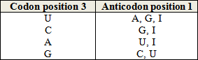
Note: I is a purine base that can pair with U, C and A but not G.
The tRNAs also have an arm of the amino acid which fixes it in 3 '(CCA) on the ribose, it is about a covalent bond: ester bond rich in energy. The amino acids will thus not arrive free on the ribosome but associated with their respective tRNAs. There are 40 to 60 different tRNAs per cell, so there are several different tRNAs for an amino acid, these are called iso-acceptor tRNAs. (Writing: tARN leu 1)
b) Loading the amino acid onto the tRNA:
The formation of the amino-acyl-tRNA complex ( aa-tRNA ) requires an amino acid - specific Amino-acyl-tRNA-synthetase , which must thus recognize all codon forms of this amino acid. Correct loading of tRNA is an important element in translational fidelity.
The amino acid (aa) is first activated and this activation requires energy in the form of ATP to allow the formation of aa-AMP (mixed anhydride bond).

The bond formed between the tRNA and the amino acid is a covalent bond of the carboxy-ester type. Amino-acyl-tRNA-synthetase is 20 in number in the cell, as many as there are amino acids that are taken into account in translation. The complexed amino acid can thus join the chain.
The specificities of eukaryotic translation
 The ribosome is of different size and composed of different ribosomal RNAs although the general structure and activity are comparable.
The initiator codon is also AUG and it is usually the 1st AUG present on the mRNA. The ATG triplet can be found which will give the AUG codon on the sense DNA at the level of the Kozak sequence (GCC GCC (A / G) CC ATG G).
As said previously, in eukaryotes the first amino acid is methionine and not the f-Met present in prokaryotes. Methionine will most often be removed just after the synthesis of the peptide chain.
The initiation factors are of the eIF (for eukaryotic initiation factor ) type, from eIF1 to eIF6.
The elongation factors are also of the EF type (EF1α, EF1β and EF2).
Termination factors are of the eRF (for eukaryotic Releasing Factor ) type.
Conclusion
The expression of the genetic information contained in DNA comprises two stages inside cells:
- transcription leading to the production of a series of ephemeral copies of the gene, the messenger RNAs;
- translation consisting of a decoding of the information contained in the messenger RNAs in the form of an assembly of amino acids. Ribosomes, globular organelles made up of two subunits, are the “assembly workshops” of proteins.